| CD83 | ||||||
|---|---|---|---|---|---|---|
| Идентификаторы | ||||||
| Символы | CD83, BL11, HB15, CD83 molecule | |||||
| Внешние IDs | MGI: 1328316 HomoloGene: 3121 GeneCards: 9308 | |||||
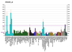
| Профиль экспрессии РНК | ||||||
 | ||||||
| Больше информации | ||||||
| Ортологи | ||||||
| Виды | Человек | Мышь | ||||
| Entrez | ||||||
| Ensembl | ||||||
| UniProt | ||||||
| RefSeq (мРНК) | ||||||
| RefSeq (белок) | ||||||
| Локус (UCSC) | Chr 6: 14.12 – 14.14 Mb | Chr 13: 43.78 – 43.8 Mb | ||||
| Поиск PubMed | ||||||
| Викиданные | ||||||
| Просмотр/Править (Человек) | Просмотр/Править (Мышь) | |||||
CD83 — мембранный белок, продукт гена человека CD83[1].
Структура
Антиген CD83 состоит из 205 аминокислот, молекулярная масса 23,0 кДа. Молекула содержит одну дисульфидную связь и 3 участка N-гликозилирования. Внеклеточный фрагмент содержит единственный иммунолглобулино-подобный домен типа V.
Функции
Может играть важную роль в презентации антигена и клеточных взаимодействиях, которые приводят к активации лимфоцитов. Наиболее характеристический маркёр полностью дифференцированных дендритных клеток[2].
Тканевая специфичность
Белок экспрессирован на активированных лимфоцитах, клетках Лангерганса и ретикулярных (дендритных) клетках паракортикальной зоны лимфатических узлов.
Примечания
- ↑ Entrez Gene: CD83 CD83 molecule.
- ↑ A. T. Prechtel, A. Steinkasserer: CD83: an update on functions and prospects of the maturation marker of dendritic cells. In: Archives of dermatological research. Band 299, Nummer 2, Mai 2007, S. 59–69, DOI:10.1007/s00403-007-0743-z, PMID 17334966 (Review).
Литература
- Lechmann M, Berchtold S, Hauber J, Steinkasserer A (2002). “CD83 on dendritic cells: more than just a marker for maturation”. Trends Immunol. 23 (6): 273—5. DOI:10.1016/S1471-4906(02)02214-7. PMID 12072358.
- Zhou LJ, Schwarting R, Smith HM, Tedder TF (1992). “A novel cell-surface molecule expressed by human interdigitating reticulum cells, Langerhans cells, and activated lymphocytes is a new member of the Ig superfamily”. J. Immunol. 149 (2): 735—42. PMID 1378080.
- Zhou LJ, Tedder TF (1995). “Human blood dendritic cells selectively express CD83, a member of the immunoglobulin superfamily”. J. Immunol. 154 (8): 3821—35. PMID 7706722.
- Maruyama K, Sugano S (1994). “Oligo-capping: a simple method to replace the cap structure of eukaryotic mRNAs with oligoribonucleotides”. Gene. 138 (1—2): 171—4. DOI:10.1016/0378-1119(94)90802-8. PMID 8125298.
- Kozlow EJ, Wilson GL, Fox CH, Kehrl JH (1993). “Subtractive cDNA cloning of a novel member of the Ig gene superfamily expressed at high levels in activated B lymphocytes”. Blood. 81 (2): 454—61. PMID 8422464.
- de la Fuente MA, Pizcueta P, Nadal M, Bosch J, Engel P (1997). “CD84 leukocyte antigen is a new member of the Ig superfamily”. Blood. 90 (6): 2398—405. PMID 9310491.
- Suzuki Y, Yoshitomo-Nakagawa K, Maruyama K, Suyama A, Sugano S (1997). “Construction and characterization of a full length-enriched and a 5'-end-enriched cDNA library”. Gene. 200 (1—2): 149—56. DOI:10.1016/S0378-1119(97)00411-3. PMID 9373149.
- Olavesen MG, Bentley E, Mason RV, Stephens RJ, Ragoussis J (1998). “Fine mapping of 39 ESTs on human chromosome 6p23-p25”. Genomics. 46 (2): 303—6. DOI:10.1006/geno.1997.5032. PMID 9417921.
- Twist CJ, Beier DR, Disteche CM, Edelhoff S, Tedder TF (1998). “The mouse Cd83 gene: structure, domain organization, and chromosome localization”. Immunogenetics. 48 (6): 383—93. DOI:10.1007/s002510050449. PMID 9799334.
- Berchtold S, Mühl-Zürbes P, Heufler C, Winklehner P, Schuler G, Steinkasserer A (1999). “Cloning, recombinant expression and biochemical characterization of the murine CD83 molecule which is specifically upregulated during dendritic cell maturation”. FEBS Lett. 461 (3): 211—6. DOI:10.1016/S0014-5793(99)01465-9. PMID 10567699.
- Scholler N, Hayden-Ledbetter M, Hellström KE, Hellström I, Ledbetter JA (2001). “CD83 is a sialic acid-binding Ig-like lectin (Siglec) adhesion receptor that binds monocytes and a subset of activated CD8+ T cells”. J. Immunol. 166 (6): 3865—72. DOI:10.4049/jimmunol.166.6.3865. PMID 11238630.
- Fanales-Belasio E, Moretti S, Nappi F, Barillari G, Micheletti F, Cafaro A, Ensoli B (2002). “Native HIV-1 Tat protein targets monocyte-derived dendritic cells and enhances their maturation, function, and antigen-specific T cell responses”. J. Immunol. 168 (1): 197—206. DOI:10.4049/jimmunol.168.1.197. PMID 11751963.
- Berchtold S, Mühl-Zürbes P, Maczek E, Golka A, Schuler G, Steinkasserer A (2003). “Cloning and characterization of the promoter region of the human CD83 gene”. Immunobiology. 205 (3): 231—46. DOI:10.1078/0171-2985-00128. PMID 12182451.
- te Velde AA, van Kooyk Y, Braat H, Hommes DW, Dellemijn TA, Slors JF, van Deventer SJ, Vyth-Dreese FA (2003). “Increased expression of DC-SIGN+IL-12+IL-18+ and CD83+IL-12-IL-18- dendritic cell populations in the colonic mucosa of patients with Crohn's disease”. Eur. J. Immunol. 33 (1): 143—51. DOI:10.1002/immu.200390017. PMID 12594843.
- Dudziak D, Kieser A, Dirmeier U, Nimmerjahn F, Berchtold S, Steinkasserer A, Marschall G, Hammerschmidt W, Laux G, Bornkamm GW (2003). “Latent Membrane Protein 1 of Epstein-Barr Virus Induces CD83 by the NF-κB Signaling Pathway”. J. Virol. 77 (15): 8290—8. DOI:10.1128/JVI.77.15.8290-8298.2003. PMC 165234. PMID 12857898.
- Hock BD, Haring LF, Steinkasserer A, Taylor KG, Patton WN, McKenzie JL (2004). “The soluble form of CD83 is present at elevated levels in a number of hematological malignancies”. Leuk. Res. 28 (3): 237—41. DOI:10.1016/S0145-2126(03)00255-8. PMID 14687618.
- Sénéchal B, Boruchov AM, Reagan JL, Hart DN, Young JW (2004). “Infection of mature monocyte-derived dendritic cells with human cytomegalovirus inhibits stimulation of T-cell proliferation via the release of soluble CD83”. Blood. 103 (11): 4207—15. DOI:10.1182/blood-2003-12-4350. PMID 14962896.
- Cao W, Lee SH, Lu J (2005). “CD83 is preformed inside monocytes, macrophages and dendritic cells, but it is only stably expressed on activated dendritic cells”. Biochem. J. 385 (Pt 1): 85—93. DOI:10.1042/BJ20040741. PMC 1134676. PMID 15320871.
Данная страница на сайте WikiSort.ru содержит текст со страницы сайта "Википедия".
Если Вы хотите её отредактировать, то можете сделать это на странице редактирования в Википедии.
Если сделанные Вами правки не будут кем-нибудь удалены, то через несколько дней они появятся на сайте WikiSort.ru .